Clinical Key Takeaways (At a Glance)
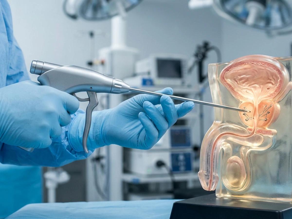
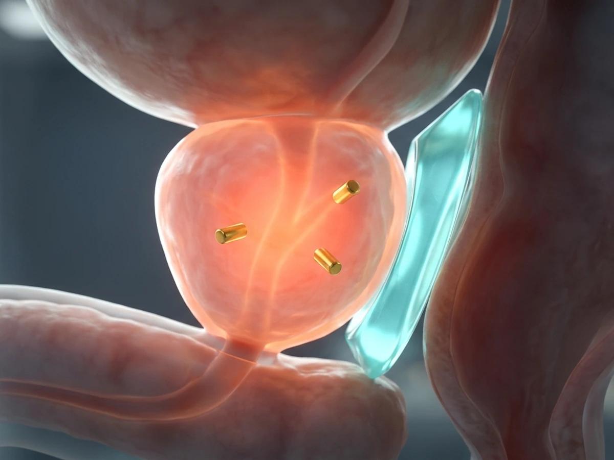
- The 1–2 mm Challenge: The anterior rectal wall lies only 1–2 mm behind the prostate, exposing it to high-dose scatter during prostate radiotherapy.
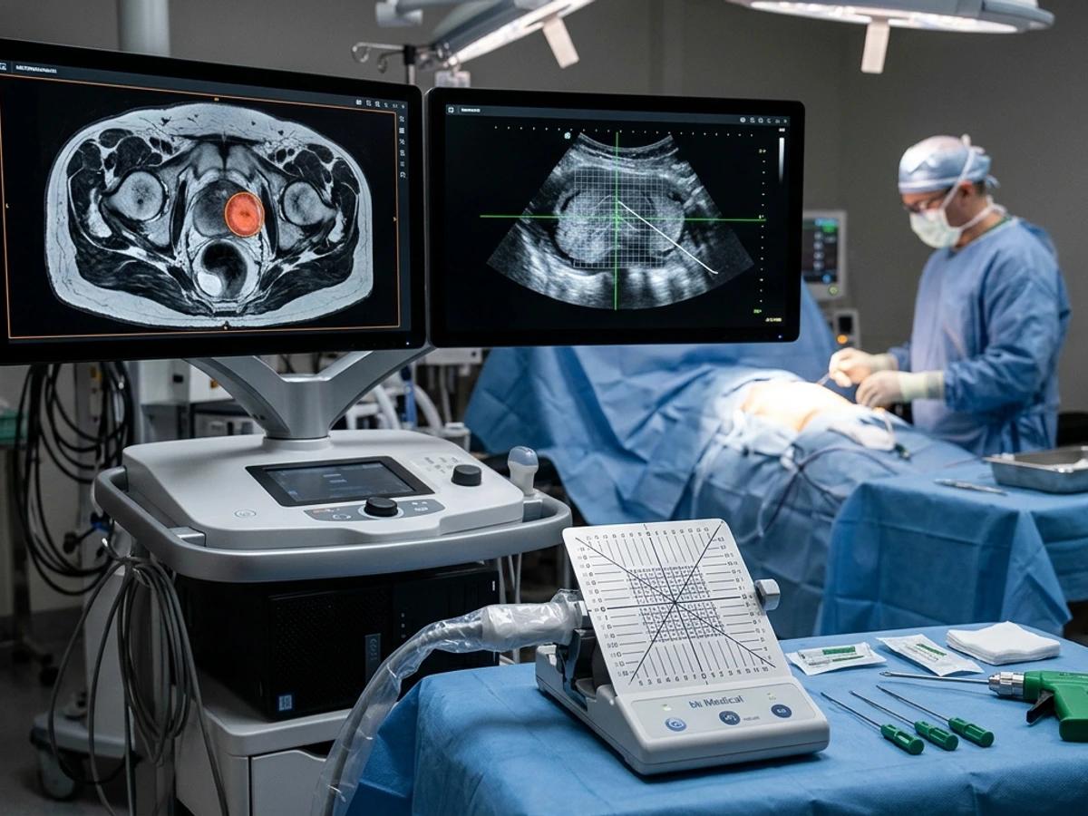
- Single Day-Case Transperineal Session: Under IV sedation and real-time side-fire TRUS, 3 gold fiducial markers (base, mid-gland, apex) are placed first, followed by saline hydrodissection and 10 mL perirectal hydrogel spacer injection.
- Level-1 Randomized Trial Proof: Pivotal trials (Mariados et al. 2015; Hamstra et al. 2017) demonstrate ~1 cm prostate-rectal separation, significantly reducing rectal radiation dose and long-term bowel toxicity.
Modern external-beam and stereotactic radiotherapy cure prostate cancer at high rates, yet the anterior wall of the rectum sits only 1 to 2 millimeters behind the prostate capsule. In this technical and patient education walkthrough, Dr. Niti Navanimitkul (ว.29673) demonstrates how combining three intraprostatic gold fiducial markers with a 10 mL absorbable rectal hydrogel spacer in a single sterile transperineal day-case session maximizes targeting accuracy while shielding the bowel.
Step-by-Step Transperineal Technique Under Side-Fire TRUS
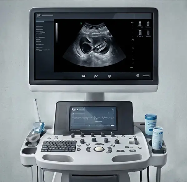
- Positioning & Probe Pressure Control: In lithotomy position under IV sedation, the side-fire transrectal ultrasound (TRUS) probe is mounted on a stepper and lowered slightly so it does not compress the potential perirectal space.
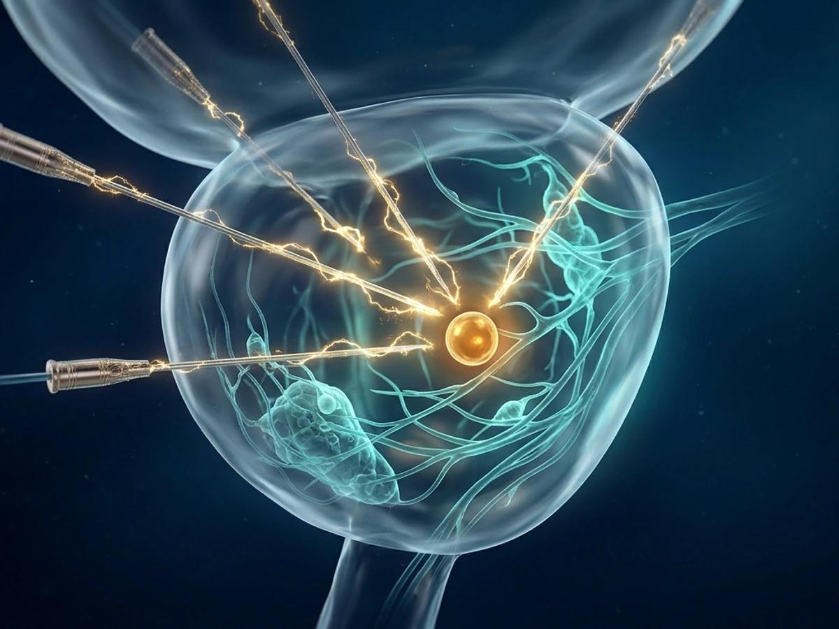
- Gold Fiducials Always First: Using preloaded needles, three 24K gold seeds are deployed asymmetrically across the prostate base, mid-gland, and apex to enable 3D daily image-guided radiotherapy tracking.
- Entering Denonvilliers' Space: An 18G needle enters on the midline 1–2 cm anterior to the anal verge with the bevel facing downward into the fat plane between Denonvilliers' fascia and the anterior rectal wall.
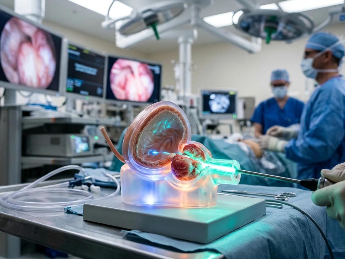
- Aspiration, Saline Hydrodissection & 10 mL Gel Push: After negative blood aspiration, sterile saline opens the space symmetrically. Then 10 mL (5 + 5 mL) of liquid hydrogel is injected smoothly in one continuous 20-second push before polymerization—while strictly monitoring for four immediate stop signals (intravascular entry, rectal wall tenting/mucosal invasion, intraprostatic tracking, or high resistance).
What Happens After Placement?
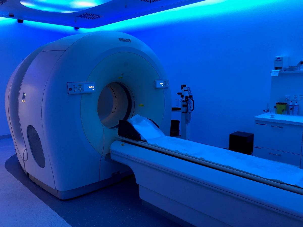
Patients go home the same day and undergo their radiotherapy planning CT and MRI scans shortly afterwards. The soft gel cushion maintains roughly 10–12 mm of protective clearance for ~3 months during radiation delivery and naturally absorbs within 6 months—avoiding spacer placement only when posterior extraprostatic tumor extension makes plane separation unsafe. Contact M-Trust Urology Clinic Pattaya to coordinate pre-radiotherapy fiducial and hydrogel spacer placement with Dr. Niti.